Introduction to Cell Biology
Relating to work conducted from 2008 to 2011
 All living things are made of cells. Cells can take on a vast variety of shapes, functions and appearances; they can live by themselves, or can function as part of a greater being. Understanding the full workings of single cells represents some of the most interesting and challenging research areas left to science.
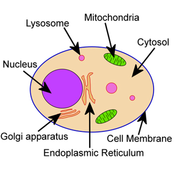
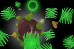
The figure, left, shows a representation of the basic components of an animal cell. Plant cells and single cell organisms are a little different, although most of these basics are the same. Around the edge of the cell is a thin membrane, separating the cell apparatus from the outside world. All over the membrane are different proteins, allowing material to be moved in and out of the cell in a controlled fashion. This allows the cell can gather food, remove waste and communicate with the outside world. When food is moved into the cell it is done via lyosomes, which are small vesicles that contain acids needed to break down the extra-cellular material. Inside the membrane is the cytosol, this is the fluid inside the cell and is filled with the material the cell needs to live. At the cell's centre is the nucleus, this contains all of the cell's DNA, the instructions needed to construct all of the other components of the cell; this is sealed off from the rest of the cell by another membrane. Outside this the endoplasmic reticulum (often known just as ER) operates to convert the instructions on the DNA into proteins that are then used to make up everything in the cell. The ER also produces the molecules needed for the membranes. The golgi apparatus is used after the ER to organize all the proteins and send them to their correct destinations. None of these processes can happen without energy, and the mitochondria are used to power the cell by converting oxygen and food into CO2 and energy.
All living things are made of cells. Cells can take on a vast variety of shapes, functions and appearances; they can live by themselves, or can function as part of a greater being. Understanding the full workings of single cells represents some of the most interesting and challenging research areas left to science.
The figure, left, shows a representation of the basic components of an animal cell. Plant cells and single cell organisms are a little different, although most of these basics are the same. Around the edge of the cell is a thin membrane, separating the cell apparatus from the outside world. All over the membrane are different proteins, allowing material to be moved in and out of the cell in a controlled fashion. This allows the cell can gather food, remove waste and communicate with the outside world. When food is moved into the cell it is done via lyosomes, which are small vesicles that contain acids needed to break down the extra-cellular material. Inside the membrane is the cytosol, this is the fluid inside the cell and is filled with the material the cell needs to live. At the cell's centre is the nucleus, this contains all of the cell's DNA, the instructions needed to construct all of the other components of the cell; this is sealed off from the rest of the cell by another membrane. Outside this the endoplasmic reticulum (often known just as ER) operates to convert the instructions on the DNA into proteins that are then used to make up everything in the cell. The ER also produces the molecules needed for the membranes. The golgi apparatus is used after the ER to organize all the proteins and send them to their correct destinations. None of these processes can happen without energy, and the mitochondria are used to power the cell by converting oxygen and food into CO2 and energy.
 Life began through something similar to a small piece of DNA being able to reproduce itself. This is not easy and requires many proteins, chemicals and reactions. Since then, evolution has refined the process of DNA replication to the point where living things exist. However, the core value of cells is still to generate DNA and to build the components needed to make this process as efficient as possible. In pursuit of this, cells have created the complex components mentioned above, and many more. It is estimated that there are over 2 million different proteins in the human body, all produced by our cells and all part of one or more processes that control some aspect of our lives. The act of a cell moving some sugar into the cytosol involves a long chain of protein interactions, all of which require more interactions to make, fold and check the proteins, and these processes also required sets of similar interactions. And so, the study of cell biology is difficult, first to see what is happing, and then to understand all the steps and proteins involved, and then to try to understand how the proteins were able to perform the tasks and work cooperatively with many others to achieve the end result. This is why cell biology is complex and interesting.
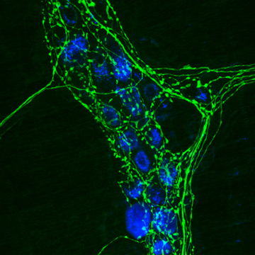
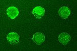
A big step in the understand of cells was the ability to stain or tag specific parts of the cell with dyes. The pioneer of this work was Antonie Philips van Leeuwenhoek. Born in 1632 Antonie discovered that saffron could be used to dye muscle fibres allowing him to observe the fibre structures more easily. For a long time, different chemicals and materials were used to see what they stained and gradually a large library of dyes was built up. This allowed great progress for many years however the biggest step was the formation of immunohistochemistry. The idea behind immunohistochemistry is that you can specifically label proteins (known as antigens) using antibodies. Antibodies are found in the blood and are used by the immune system to identify foreign objects. In immunohistochemistry the antibody for a specific protein must first be found then cultured with the cell. Antibodies bind to their specific targets, so attach to the proteins of interest, then in a second step these antibodies are made to be fluorescent by using a second protein with a dye that will attach to the first (and only the first) protein. While complicated, this process has been very successful to greatly extend the knowledge of where specific proteins are located in cells and how they move when the cell is subjected to different stimuli. The image, copied form Wikipedia, shows a beautiful example of the labelling of a protein involved in the production of adrenaline in a cell involved with the nervous system. This clearly shows what is where the proteins are across the cell and if watch over time it becomes possible to see trends and gain better understanding of what the cell is doing.
In the work we have been undertaking the aim is to replace the dye in the above example with a nano-particle. There are several reasons to do this, first nano-particles are brighter and last longer inside a cell than dye molecules. Secondly a nano-particle can have multiple peptides attached to it, so as to enhance its interactions with cell, be useful as a miniature drug delivery capsule, and be possible to have a number of different functions when a variety of peptides are present. As steps to understanding these goals we have first investigated the endocytosis of nano-particles, and then developed the necessary proteins to study receptor mediated endocyctosis of a peptide of interest - somatostatin, a protein which helps regulates blood pressure. We have then developed the necessary chemistry to attach peptides to nano-particles of interest using a protein linker pair.
Papers relating to this work:
 Non-specific cellular uptake of surface-functionalized quantum dots
T.A. Kelf, V.K.A. Sreenivasan, J. Sun, E.J. Kim, E.M. Goldys and A.V. Zvyagin
Nanotechnology.21, 285105 (2010)
 Pharmacological Characterization of a Recombinant, Fluorescent Somatostatin Receptor Agonist
V.K.A. Sreenivasan, O.A. Stremovskiy, T.A. Kelf, M. Heblinski, A.K. Goodchild, M. Connor, S.M. Deyev, and A.V. Zvyagin
Bioconjugate. Chem. 22, 1768-1775 (2011)
 Barstar:barnase - a versatile platform for colloidal diamond bioconjugation
V.K. . Sreenivasan, E.A. Ivukina, W. Deng, T.A. Kelf, T.A. Zdobnova, S.V. Lukash, B.V. Veryugin, O.A. Stremovskiy, A.V. Zvyagin, and S.M. Deyev
J. Mat. Chem.21, 65 (2011)
 A modular design of low-background bioassays based on a high-affinity molecular pair barstar:barnase
V.K.A. Sreenivasan, T.A. Kelf, E.A. Grebenik, O.A. Stremovskiy, J.M. Say, J.R. Rabeau, A.V. Zvyagin, S.M. Deyev.
Proteomics. 13, 1737-1443 (2013)